Managing trauma in exotic animals requires a careful balance between clinical effectiveness and minimal invasiveness. In reptiles, and particularly in chelonians, tissue fragility, slow healing processes, and anesthetic risks make trauma cases especially complex.
This case involves a Chinese stripe-necked turtle (Mauremys sinensis) presented in emergency conditions after a traumatic fall. Thanks to a conservative therapeutic approach supported by radiofrequency technology, it was possible to stabilize the fracture, promote tissue healing, and avoid surgical intervention.
We asked Dr. Valentina Bruson to share the case, her therapeutic choices, and how this technology supports trauma management in exotic animals.
I graduated in 2006 and soon began working both independently and in a small animal clinic in the Canavese area (Northern Italy). At the same time, I trained at Dr. Mattia Bielli’s practice in Novara (Northern Italy), where I developed strong expertise in exotic animal medicine. In 2010, I opened my own clinic, expanding it in 2015 with a laboratory, surgical room, and short-term hospitalization area. We mainly treat exotic pets and wildlife, following a holistic medical approach and carefully limiting the excessive use of drugs.
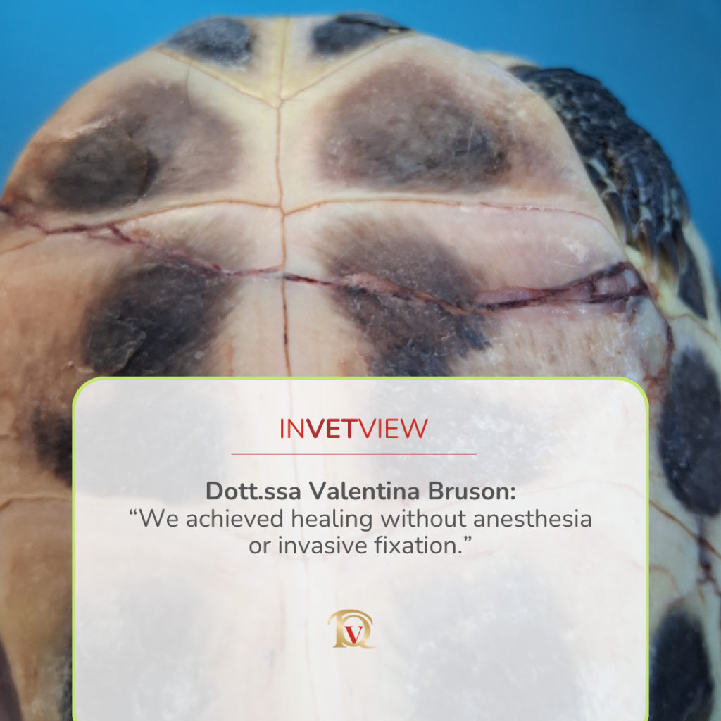
The turtle, later named Maura, was brought to the clinic after falling from a second-floor balcony. On clinical examination, the animal appeared alert and responsive, with preserved mobility of the limbs and head. However, two significant shell fractures were immediately evident: a complete fracture of the plastron, characterized by instability and mobility of the bone fragment and a partial fracture of the carapace, involving only the superficial layers. Radiographic evaluation confirmed the presence of the plastron fracture and revealed mild pulmonary contusion, likely caused by the impact of the fall. Given the extent of the injuries, the primary clinical concerns were pain management, prevention of infection, stabilization of the fracture margins, and preservation of respiratory function.
Although shell fractures may appear similar to fractures in other species, they involve living bone tissue that is richly innervated and highly sensitive. These injuries are therefore extremely painful and prone to contamination, especially in aquatic or semi-aquatic species. In turtles, bone and shell healing is notoriously slow and may take several months or even more than a year. During this period, any instability or infection can severely compromise the outcome. Additionally, open shell fractures prevent normal water access, which is essential for hydration, feeding behavior, and overall well-being in semi-aquatic turtles. This significantly impacts the animal’s quality of life and complicates long-term management.
The first priority is always to restore the reptile’s metabolism to the most optimal condition possible; without this, the patient will not respond adequately to any pharmacological treatment. For this reason, Maura was immediately placed in a heated enclosure maintained at 24–25°C. Water was intentionally excluded, while a moist towel was provided to allow adequate humidity without compromising wound management. According to the owners, the turtle had experienced some blood loss following the fall. For this reason, subcutaneous fluid therapy was administered to restore hydration. Subsequently, parenteral medications were initiated, including: meloxicam for pain and inflammation control, arnica to support tissue recovery and enrofloxacin as antibiotic coverage. In addition to conventional medical treatment, I routinely provide Reiki sessions to hospitalized patients. In this case, Reiki was used primarily to help rebalance the animal following the traumatic experience of the fall, supporting overall recovery and stress management.
Radiofrequency treatment was introduced two days after the traumatic event. Although the wound appeared externally clean and it would have been possible to intervene immediately, the patient first needed to be stabilized. I also preferred to allow the antibiotic therapy to begin taking effect. Even when a wound looks clean from the outside, in cases where several hours have passed since the traumatic event, it is always reasonable to assume some degree of bacterial penetration. Since my objective was to perform a biostimulatory treatment, timing was crucial. The primary goal was to accelerate fracture healing as much as possible, in order to allow the animal to return to a normal life including access to water in the shortest possible time.
Radiofrequency technology promotes local vascularization and activates various chemotactic factors, stimulating the repair of damaged tissues both soft tissues and hard tissues. In patients where complete stabilization and healing of a fracture may require many months, often with significant management challenges for the owners, having a tool that can help shorten recovery time becomes extremely valuable. Additionally, radiofrequency promotes the recruitment of immune system cells, thereby reducing the risk of microbial spread and secondary infections.
I used radiofrequency with a bipolar resistive transducer at 1000 MHz, as I was working on a highly resistive but superficial bone tissue. The impulse duration was set low (250) with a long pause (750) to achieve a biostimulatory effect.Power was set at 13%, allowing me to work in a near athermic mode, while still considering the solidity of the tissue to be penetrated. The handpiece was used in combination with ArniTop, a conductive cream containing arnica, a plant known for its anti-inflammatory and wound-healing properties.
After the first session, the fracture margins already appeared closer to one another, despite the absence of any additional fixation. After the second treatment, the previously mobile fragment was stable and well aligned, and the turtle was able to take a first short bath lasting a few minutes.
No, I did not use any additional fixation systems. This allowed me to avoid anesthesia, which would have been necessary for fixation techniques such as cerclage wiring. It also meant avoiding the application of glues or resins which, although capable of providing stabilization, must be applied with extreme precision to avoid worsening the lesion. Moreover, these materials are not breathable and do not allow for further inspection or local wound management.
The main advantage is undoubtedly the ability to treat the patient without anesthesia. Anesthesia in reptiles is not always straightforward: despite advances in veterinary medicine, drug responses are not always precise or predictable, particularly in patients that often suffer from stress and/or chronic subclinical conditions.
In this specific case, such procedures were fortunately not necessary. However, there are lesions that may require multiple curettage surgeries to remove devitalized tissue, such as bite wounds. In these cases, after an initial thorough wound cleaning, I now apply radiofrequency immediately. This approach has allowed me to revitalize surrounding tissues and, in a couple of cases, even to avoid limb amputation.
It allows us to offer caregivers a valid, non-invasive alternative, applicable immediately or almost immediately, and one that enables regular patient monitoring and follow-up. Furthermore, even when surgery is ultimately required, this technique often allows me to delay the procedure until the patient is better stabilized, or when applied postoperatively to significantly accelerate healing.
Certainly the complete absence of mobility of the fractured fragment after only two treatment sessions.I am accustomed to seeing faster wound healing since I started using radiofrequency, but this particular case progressed remarkably quickly.
Absolutely, I would recommend it. To be honest, I now use radiofrequency on almost everything. It is highly effective in cases of fractures or traumatic injuries in general, as well as post-surgery, to promote faster healing. It is worth remembering that even a surgical wound, although planned, is still a wound.
Yes. In the past, I would immediately schedule surgery regardless of the circumstances.
Now, this is no longer the case. I have a technique that allows me to resolve many situations without anesthetizing the patient, and even when that is not sufficient, it gives me more time to address other underlying clinical or management issues which are very common in reptiles and, last but not least, to achieve better owner compliance.
Dr. Bruson, thank you for sharing this clinical case and for clearly and rigorously describing your approach to managing such a delicate situation. Your experience highlights how, in modern veterinary medicine, professional expertise and the conscious selection of therapeutic tools can truly make a difference in the healing process.
Copyright © Top Quality Group S.r.l.
Companies Register of Perugia No. / Tax Code / VAT Number 03424560542
REA PG No. 288065
Fill in the fields below to receive the complete technical data sheet of the device directly in your email.
Compila i campi sottostanti per ricevere direttamente nella tua email la scheda tecnica completa del dispositivo.